41 drawing of compound microscope with label
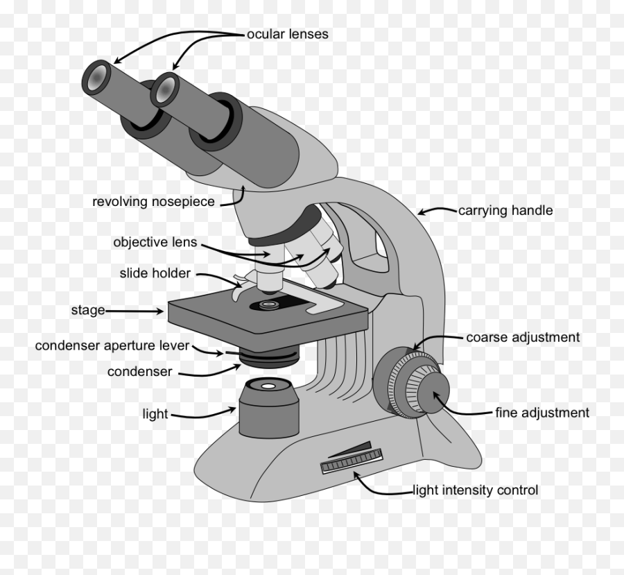
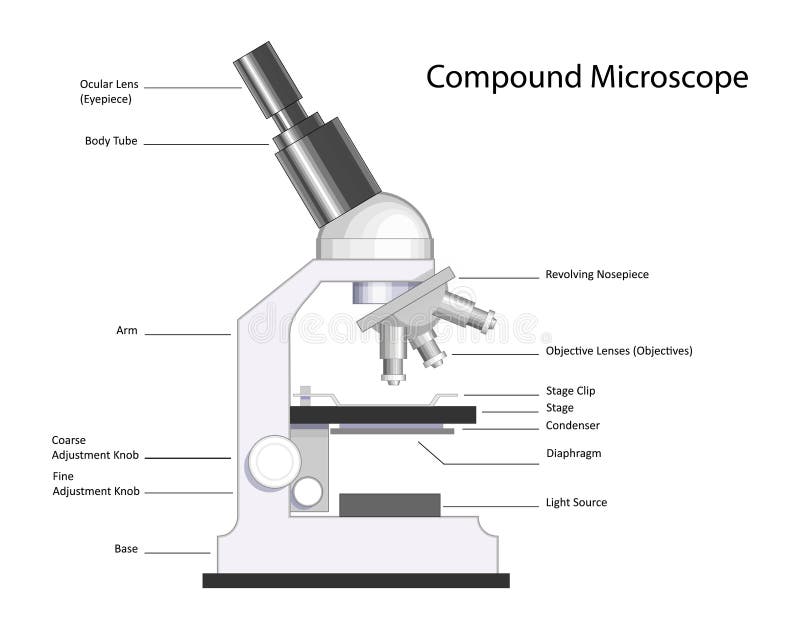
Microscope Parts and Functions With Labeled Diagram and Functions How does a Compound Microscope Work? Before exploring microscope parts and functions, you should probably understand that the compound light microscope is more complicated than just a microscope with more than one lens. Compound Microscope Parts - Labeled Diagram and their Functions Labeled diagram of a compound microscope Major structural parts of a compound microscope There are three major structural parts of a compound microscope. The head includes the upper part of the microscope, which houses the most critical optical components, and the eyepiece tube of the microscope.
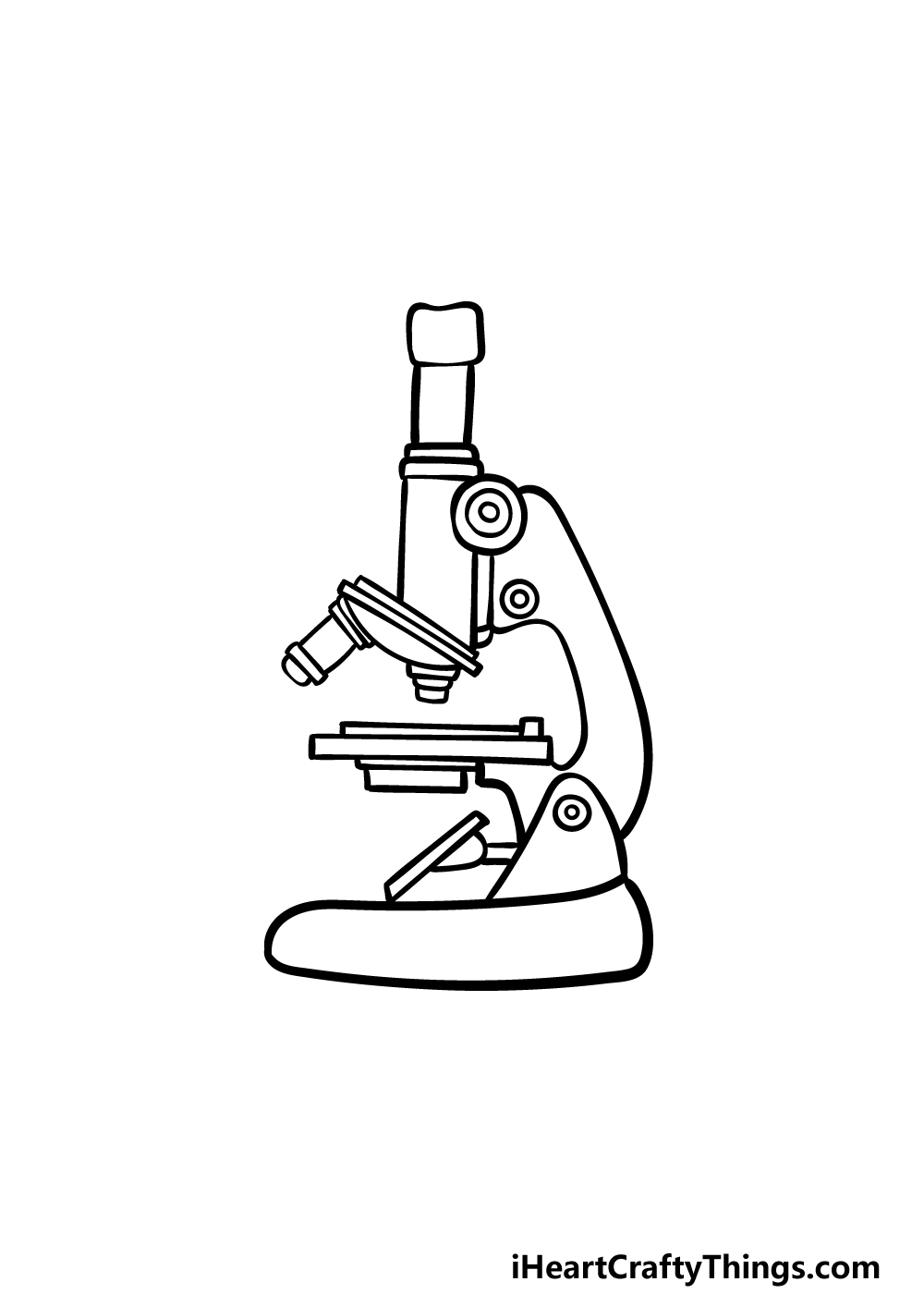
How to Draw a Microscope - Realistic Microscope Drawing Tutorial Step 3: Outline the Arm Frame. We are now going to draw the arm that the microscope uses to swivel back and forth. Attached to the right side of the head from the previous step, draw two curving lines. You can then make this three-dimensional by drawing two smaller curved lines in the way you can see below.

Drawing of compound microscope with label
Label the microscope — Science Learning Hub In this interactive, you can label the different parts of a microscope. Use this with the Microscope parts activity to help students identify and label the main parts of a microscope and then describe their functions. Drag and drop the text labels onto the microscope diagram. How to draw compound of Microscope easily - step by step How to draw compound of Microscope easily - step by step Perhaps Bidesh 52.4K subscribers Subscribe 1.4M views 3 years ago Biology diagram I will show you " How to draw compound of microscope... Drawing Of A Compound Microscope - Warehouse of Ideas Drawing Of A Compound Microscope. Add answer + 15 pts. At the side of the microscope place a sheet of white drawing paper with a pencil on, lying beneath the mirror. diagram of compound microscope Brainly.in from brainly.in. Copperplate engraving by wilson lowry after a drawing by j. Body inclined at 45º for fatigue free viewing can be rotated ...
Drawing of compound microscope with label. Draw a labelled diagram of an image formed by a compound microscope ... Draw a labelled diagram of an image formed by a compound microscope, with the image at least distance of distinct vision. Write any one expression for its magnifying power. Medium Solution Verified by Toppr Expression of magnifying power of a compound microscope is given by: m=− u ov o(1+ f eD) A Study of the Microscope and its Functions With a Labeled Diagram ... These labeled microscope diagrams and the functions of its various parts, attempt to simplify the microscope for you. However, as the saying goes, 'practice makes perfect', here is a blank compound microscope diagram and blank electron microscope diagram to label. Labeling the Parts of the Microscope | Microscope World Resources Each microscope layout (both blank and the version with answers) are available as PDF downloads. You can view a more in-depth review of each part of the microscope here. Download the Label the Parts of the Microscope PDF printable version here. Download the Label the Parts of the Microscope: Answers PDF printable version here. Diagram of a Compound Microscope - Biology Discussion 1. It is noted first that which objective lens is in use on the microscope. 2. Stage micrometer is positioned in such a way that it is in the field of view. 3. The eyepiece is rotated so that the two scales, the eyepiece or ocular scale and the stage micrometer scale, are parallel. 4.
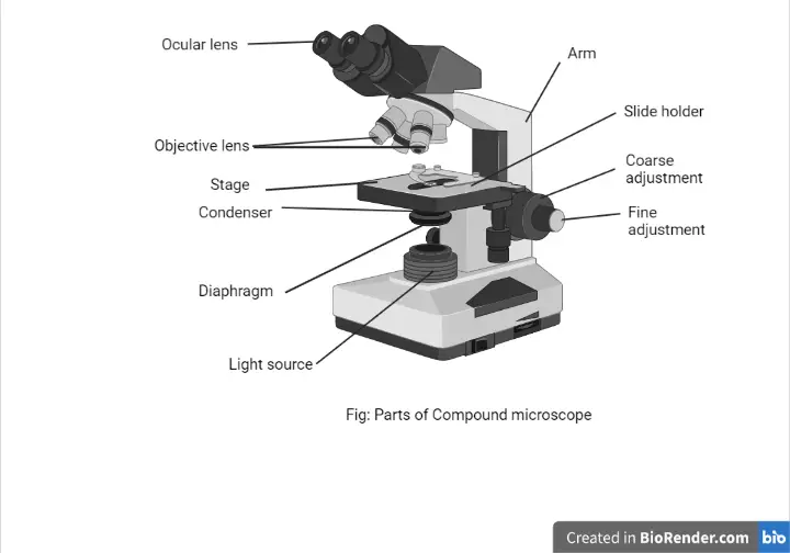
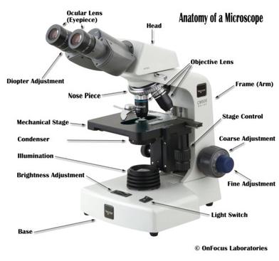
(b) Why both objective and eyepiece of a compound microscope must have ... (a) Draw the labelled ray diagram for the formation of image by a compound microscope. Derive an expression for its total magnification (or magnifying power), when the final image is formed at the near point. (b) Why both objective and eyepiece of a compound microscope must have short focal lengths? Parts of a microscope with functions and labeled diagram - Microbe Notes Head - This is also known as the body. It carries the optical parts in the upper part of the microscope. Base - It acts as microscopes support. It also carries microscopic illuminators. Arms - This is the part connecting the base and to the head and the eyepiece tube to the base of the microscope. How to Draw a Microscope - Really Easy Drawing Tutorial Easy Microscope Drawing - Step 2 2. Extend a pair of straight, parallel lines from the head, and connect them at the end using a short curved line. This forms the eyepiece tube. The tip of the tube is called the ocular. It is the lens through which you look to view your tiny objects. Easy Microscope Drawing - Step 3 3. Compound Microscope - Types, Parts, Diagram, Functions and Uses It comes with a wide body and base. Its distinct parts include a condenser, illumination, focus lock, mechanical stage, and a revolving nosepiece which can hold up to five objectives. It usually has a binocular head, which makes long-term observation easy. Image 22: An example of a research compound microscope.
Compound Microscope: Definition, Diagram, Parts, Uses, Working ... - BYJUS A compound microscope is defined as A microscope with a high resolution and uses two sets of lenses providing a 2-dimensional image of the sample. The term compound refers to the usage of more than one lens in the microscope. Also, the compound microscope is one of the types of optical microscopes. Parts of the Microscope with Labeling (also Free Printouts) Microscopes are specially created to magnify the image of the subject being studied. This exercise is created to be used in homes and schools. the microscope layout, including the blank and answered versions are available as pdf downloads. Click to Download : Label the Parts of the Microscope (A4) PDF print version. Drawing Of A Microscope And Label - Warehouse of Ideas Here presented 54+ microscope drawing and label images for free to download, print or share. Title Is Informative, Centered, And Larger Than Other Text. How to draw a microscope and label. Compound microscopes have furthered medical research, helped to solve crimes, and they have repeatedly proven invaluable in unlocking the secrets of the. Compound Microscope Drawing - Painting Valley We collected 37+ Compound Microscope Drawing paintings in our online museum of paintings - PaintingValley.com. ADVERTISEMENT. Most Downloads Size Popular. Views: 11196 Images: 37 Downloads: 190 Likes: 17. microscope; compound; ... Microscope Drawing And Label. Microscope Drawing Worksheet.
Microscope Labeling
(a) Draw a labelled ray diagram of a compound microscope. (b) Derive an ... (a) Labelled diagram of compound microscope. The objective lens form image A' B' near the first focal point of eyepiece. (b) Angular magnification of objective lens m 0 = linear magnification h'/h. where L is the distance between second focal point of the objective and first focal point of eyepiece.If the final image A'' B'' is formed at the near point.

Draw a well labelled diagram of a microscope. - Brainly.in
Compound Microscope- Definition, Labeled Diagram, Principle, Parts, Uses A compound microscope is of great use in pathology labs so as to identify diseases. Various crime cases are detected and solved by drawing out human cells and examining them under the microscope in forensic laboratories. The presence or absence of minerals and the presence of metals can be identified using compound microscopes.

Lasec Education | Key parts of a compound microscope and how ...
Microscope Drawing Easy with Label - YouTube Apr 13, 2020 3 Dislike Share DrDiclonius 1.76K subscribers In this video I go over a microscope drawing that is easy with label. There is a blank copy at the end of the video to review on...
Microscopy Lab The Biology Primer - Drawing Compound ...
16 Parts of a Compound Microscope: Diagrams and Video The 16 core parts of a compound microscope are: Head (Body) Arm Base Eyepiece Eyepiece tube Objective lenses Revolving Nosepiece (Turret) Rack stop Coarse adjustment knobs Fine adjustment knobs Stage Stage clips Aperture Illuminator Condenser Diaphragm Video: Parts of a compound Microscope with Diagram Explained

The Compound Microscope. - ppt download
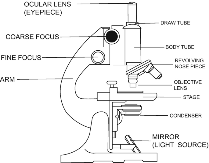
Labelled Diagram of Compound Microscope Labelled Diagram of Compound Microscope Article Shared by ADVERTISEMENTS: The below mentioned article provides a labelled diagram of compound microscope. Part # 1. The Stand: The stand is made up of a heavy foot which carries a curved inclinable limb or arm bearing the body tube.

Draw a neat labelled diagram of a compound microscope and ...
Compound Microscope: Parts of Compound Microscope - BYJUS The parts of the compound microscope can be categorized into: Mechanical parts; Optical parts (A) Mechanical Parts of a Compound Microscope. 1. Foot or base. It is a U-shaped structure and supports the entire weight of the compound microscope. 2. Pillar. It is a vertical projection. This stands by resting on the base and supports the stage. 3. Arm
Parts of a Microscope with Their Functions • Microbe Online
Drawing Of A Compound Microscope - Warehouse of Ideas Drawing Of A Compound Microscope. Add answer + 15 pts. At the side of the microscope place a sheet of white drawing paper with a pencil on, lying beneath the mirror. diagram of compound microscope Brainly.in from brainly.in. Copperplate engraving by wilson lowry after a drawing by j. Body inclined at 45º for fatigue free viewing can be rotated ...

Compound microscope and its parts:3 Diagram | Quizlet
How to draw compound of Microscope easily - step by step How to draw compound of Microscope easily - step by step Perhaps Bidesh 52.4K subscribers Subscribe 1.4M views 3 years ago Biology diagram I will show you " How to draw compound of microscope...

Labeling the Parts of the Microscope | Microscope World Resources
Label the microscope — Science Learning Hub In this interactive, you can label the different parts of a microscope. Use this with the Microscope parts activity to help students identify and label the main parts of a microscope and then describe their functions. Drag and drop the text labels onto the microscope diagram.

Can someone can send me diagram of this compound microscope ...

The compound light microscope
Exercise 1: Using a Compound Microscope | SpringerLink

HOw to draw light or compound microscope step by step / Microscope diagram
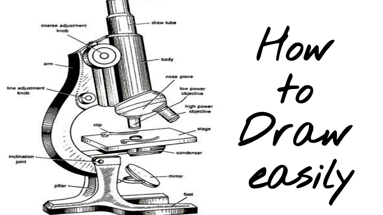
How to draw compound microscope | science apparatus | compound microscope

How to Draw a Microscope - Really Easy Drawing Tutorial
Compound Microscope Drawing - ClipArt Best
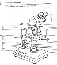
Answered: Microscope Structure and Function… | bartleby
compound-microscope-unlabeled.jpg
1.2: Microscopes - Biology LibreTexts

Compound Microscope: Parts of Compound Microscope
Microscope Drawing - How To Draw A Microscope Step By Step

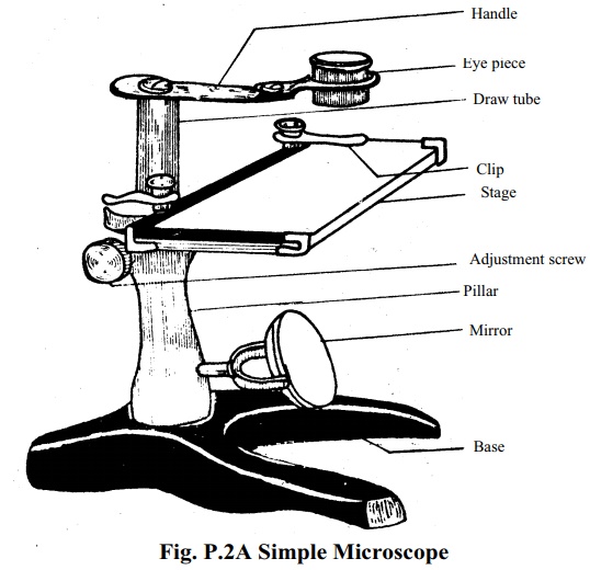
Simple Microscope - Diagram (Parts labelled), Principle ...

Remix of "The Compound Microscope"
Compound Microscope stock vector. Illustration of research ...

in a long bond paper draw and label the parts of a compound ...

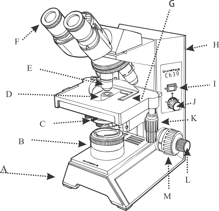
Solved A. OLYMPUS C. B. Use the Diagram to answer the | Chegg.com

Cell Drawing Microscope - Binocular Compound Microscope ...
Microscope: Structure, Uses, Functioning Processes of Simple ...
Microscope Parts and Functions
A typical microscope | Download Scientific Diagram

Microscope Diagram Labeled, Unlabeled and Blank | Parts of a ...

Compound microscope illustration - Lizzie Harper

Compound Microscope Parts, Functions, and Labeled Diagram ...

Compound Microscope – Diagram (Parts labelled), Principle and ...

Compound Microscope- Definition, Labeled Diagram, Principle ...

Microscope Parts, Types & Diagram | What is a Microscope ...

Compound microscope Diagram | Quizlet
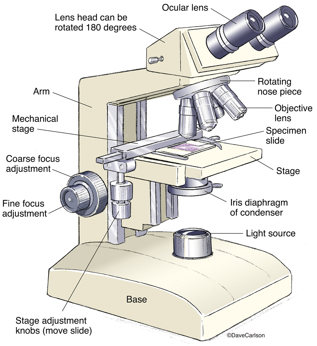
Compound Microscope | Image License | Carlson Stock Art

How to draw microscope/Draw microscope in simple way.

Simple doodles, Microscopic images, Microscope parts
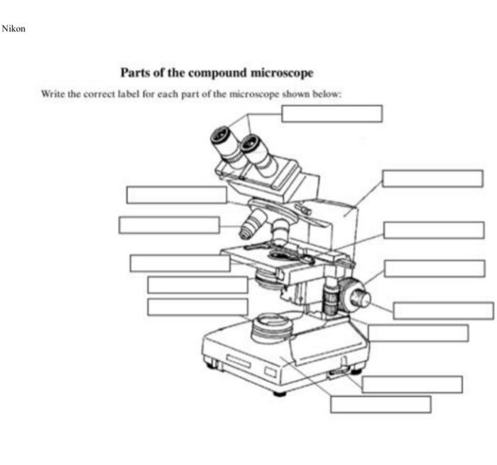
Solved Nikon Parts of the compound microscope Write the ...
Post a Comment for "41 drawing of compound microscope with label"